
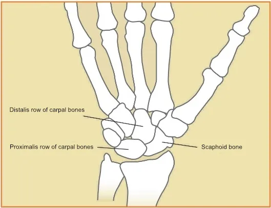
There are eight small carpal bones in the wrist, one of which is the scaphoid. These small bones are arranged in two rows: one row is closer to the forearm (the proximal row of carpal bones), and the other is closer to the fingers (the distal row of carpal bones). The scaphoid is unique in that it connects these two rows (Figure 1). This bone is the most commonly injured of the carpal bones.
Fracture of the scaphoid bone
Fracture of the scaphoid bone
Outpatient specialties | Orthopaedics
Fracture of the scaphoid bone
What is a scaphoid fracture?
How does the scaphoid bone break?
The scaphoid bone is most commonly fractured during a forward fall with an outstretched hand. Initially, pain sets in, which subsides relatively quickly over the course of a few days or weeks. Minor swelling develops; there is generally no visible deformity, and bruising occurs very rarely. Since there is no visible deformity and the pain subsides, many people assume they have simply sprained their wrist and delay seeking medical attention or do not seek it at all. Often, the fracture is not discovered until months or even years after the fall.
Recognizing a Scaphoid Fracture
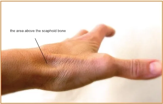
A scaphoid fracture can usually be detected on an X-ray of the wrist. However, if the fracture has not displaced, it may not always be visible on an X-ray taken early on (during the first week). Thus, a non-displaced scaphoid fracture can, unfortunately, easily be overlooked. Therefore, if marked tenderness is palpable over the scaphoid bone (Figure 2), a scaphoid fracture is suspected, and immobilization is necessary. On a repeat X-ray taken one or two weeks after the injury, the fracture is clearly visible. In uncertain cases, an MRI scan helps establish the diagnosis as soon as possible. Until a definitive diagnosis is made, wearing a cast is recommended to prevent displacement of the fracture.
Treatment of a scaphoid fracture
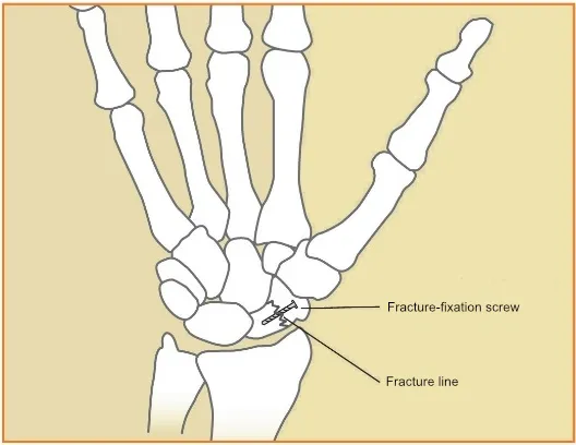
If the fracture has not shifted, a cast is required for 8–12 weeks. The fracture must be immobilized for this long because the scaphoid bone has a poor blood supply, and adequate blood flow is necessary for healing. Part of the bone may even die following a fracture if the fracture cuts off the blood supply to that part of the bone—this is particularly dangerous at the proximal pole of the radius, which is the part of the bone closest to the forearm. If the fracture occurs in this area or displacement is observed, surgery is recommended. During surgery, a screw is inserted into the bone to stabilize the fracture. (Figure 3) Occasionally, a bone graft is also necessary to aid in the healing of the fracture. To avoid prolonged immobilization, the option of fixation with a screw is sometimes considered even in cases without displacement.
Complications of scaphoid fractures
Pseudoarthrosis: If a scaphoid fracture goes unrecognized, it often fails to heal. In some cases, even with proper treatment, the fracture does not heal due to poor blood supply. Over time, the resulting abnormal movements and the reduction in the size of the broken bone fragments alter the fundamental statics and biomechanics of the wrist, leading to wear and tear in the long term. If the pseudoarthrosis is detected before wear and tear develops, surgery to restore the scaphoid bone is recommended.
Avascular necrosis: A portion of the scaphoid bone may die due to a lack of blood supply, causing the bone to shrink, which leads to wear and tear in the long term. The most at-risk area is the proximal third of the bone—that is, the half closest to the forearm. If the condition is detected before joint wear sets in, surgery is recommended to restore blood flow and heal the fracture.
Wear and tear: Once wear and tear has developed, palliative procedures are considered to reduce pain and preserve mobility. In extreme cases, partial or complete fusion of the wrist may be necessary.
© 2026 PASAMED.